Drosophila
calcium imaging of odor evoked
activity along the olfactory pathway.
This page relates to the paper Lüdke et al., 2018:
“Calcium in Kenyon Cell Somata as a Substrate for an Olfactory Sensory Memory in
Drosophila ”.
Frontiers in Cellular Neuroscience, 14 May 2018, 12:128 |
https://doi.org/10.3389/fncel.2018.00128
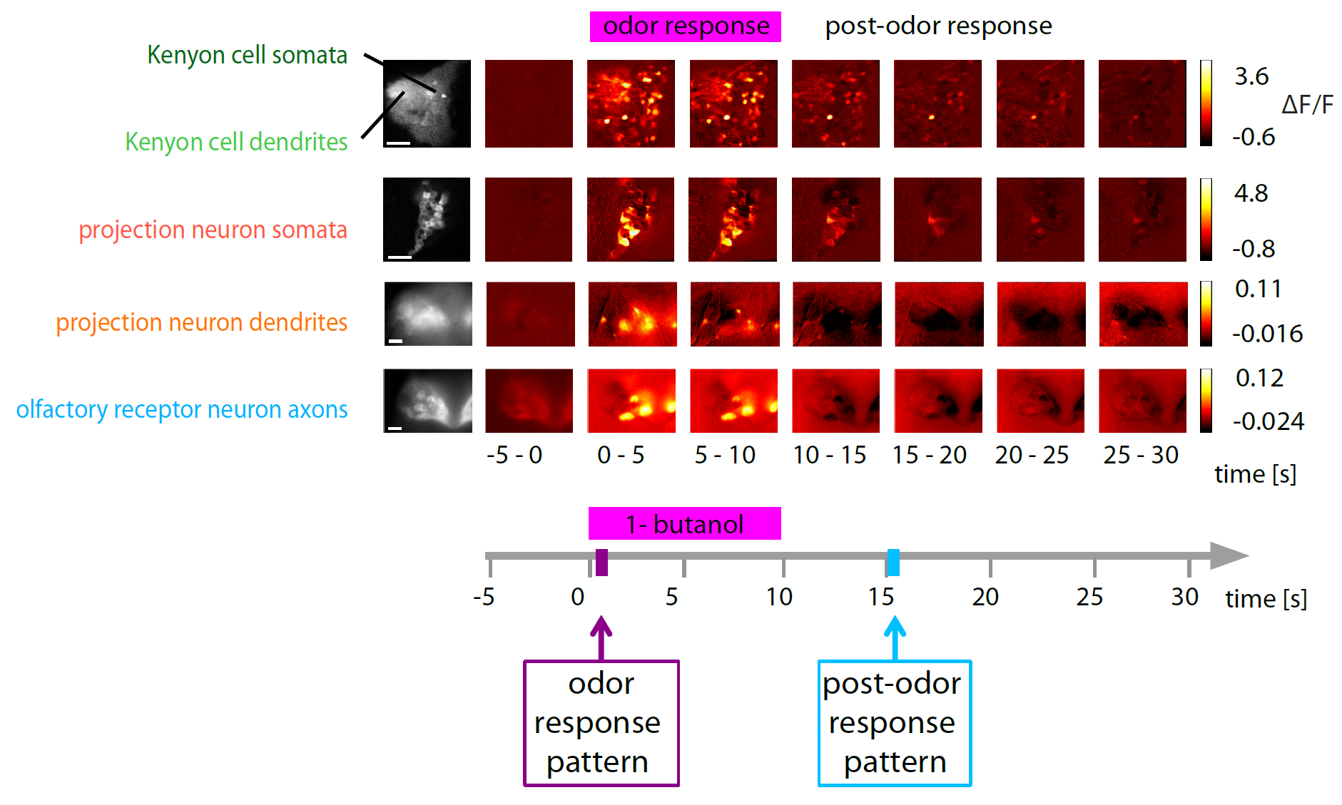
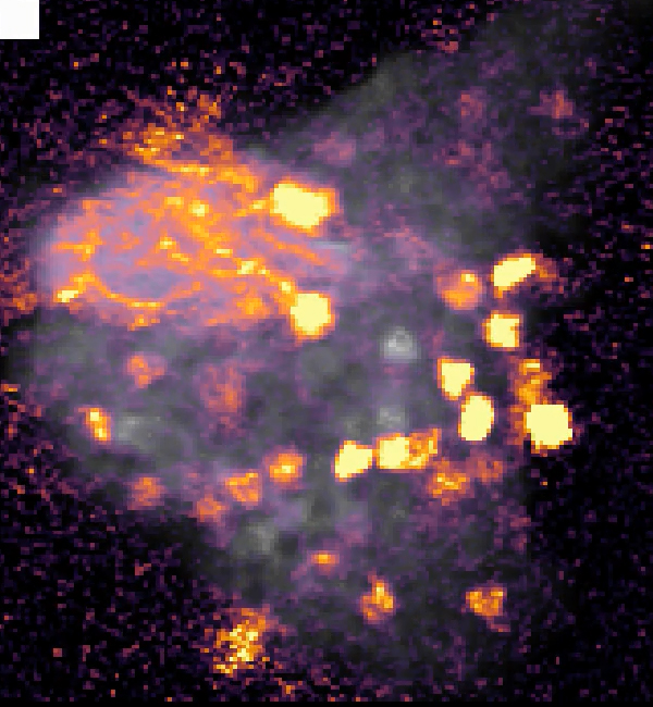
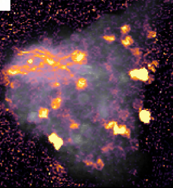
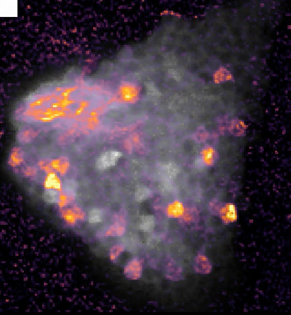
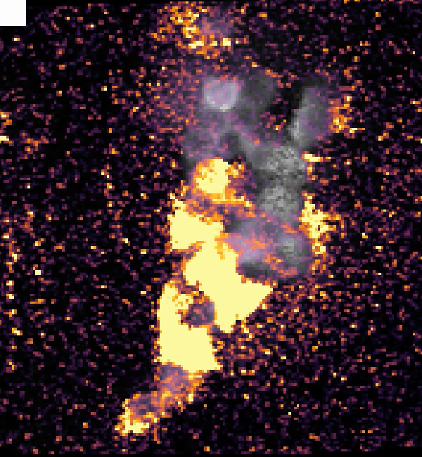
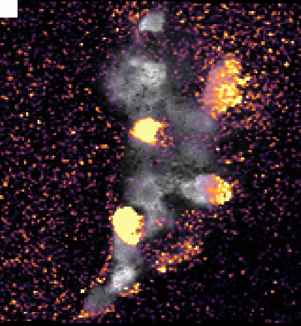
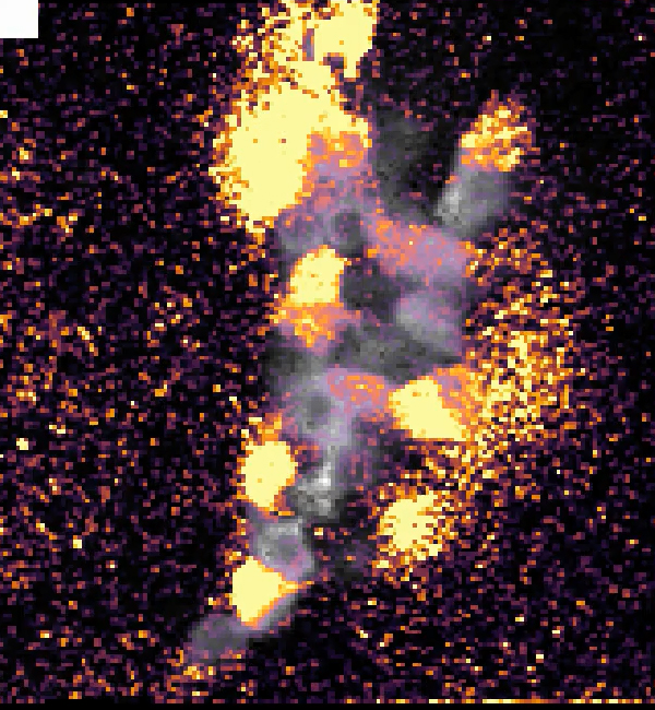
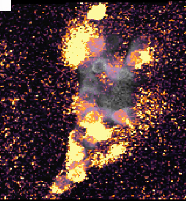
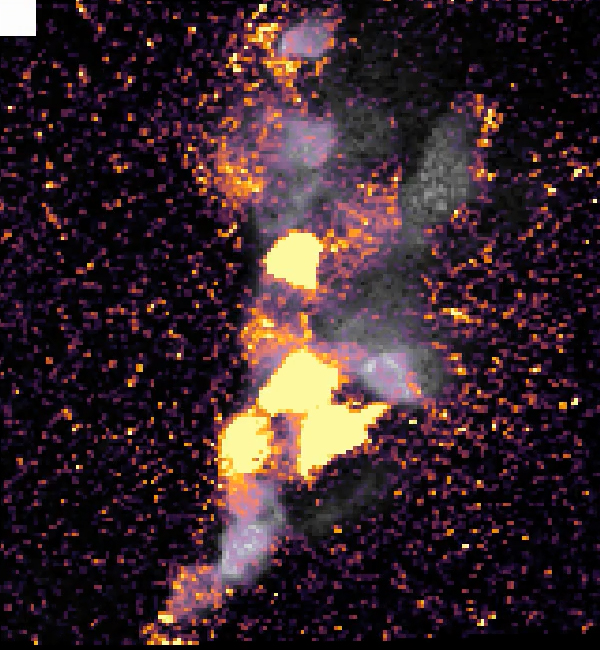
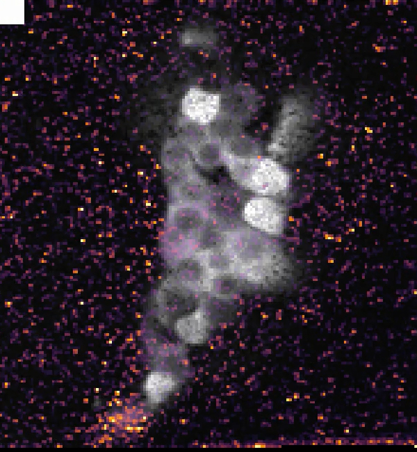
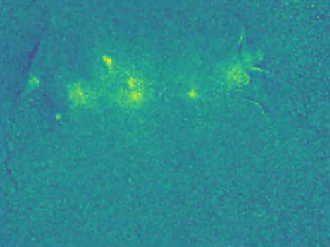
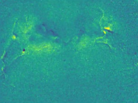
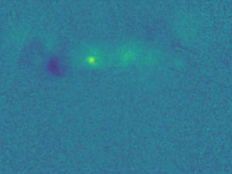
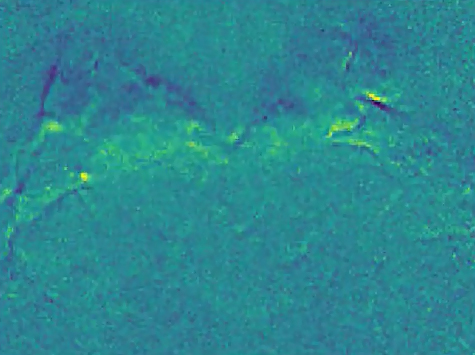
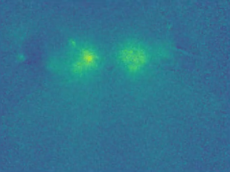
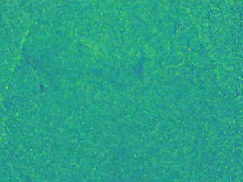
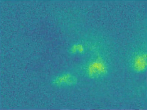
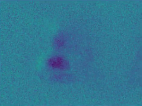
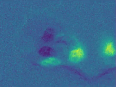
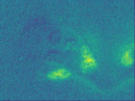
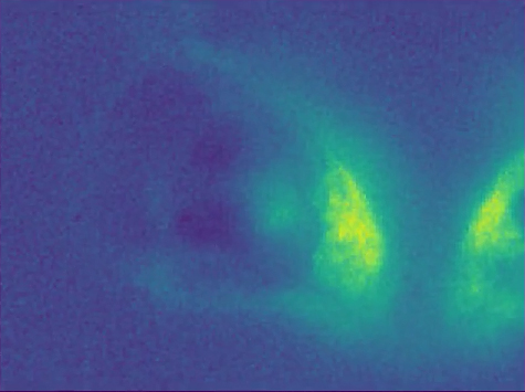
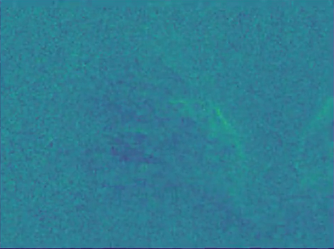
Figure 1B: Odor and post-odor responses along the olfactory pathway.
Spatial distribution of Ca2+ activity during and after olfactory stimulation (here with 1-butanol) in the neuronal compartments along the olfactory pathway.
Color-coded
DF/F images show the average of 5 s recording time (i.e., 25 frames).
All areas showed distinct responses upon odorant stimulation. (Scale bars 20 µm).
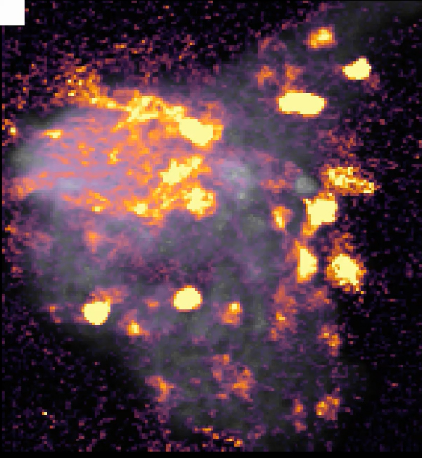
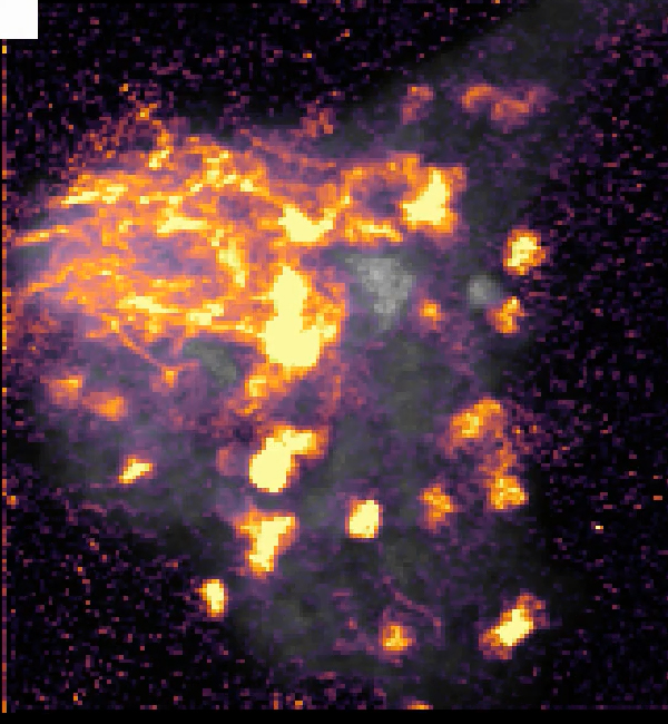
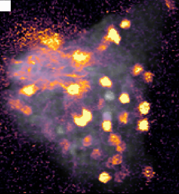
Below, see the calcium imaging movies with different odorant stimulations.
The movies are 35 s long, odorant stimulation starts after 5 s and lasts for 10 s (indicated by a white square in the upper left corner).
| |
1-butanol |
acetic acid |
propanoic acid |
1-propanol |
4-methyl-cyclohexanol |
mineral oil |
| KC somata & dendrites |
 |
 |
 |
 |
 |
 |
| |
|
|
|
|
|
|
| PN somata |
 |
 |
 |
 |
 |
 |
| |
|
|
|
|
|
|
| |
1-butanol |
acetic acid |
propanoic acid |
1-propanol |
ethyl acetate |
mineral oil |
| PN dendrites |
 |
 |
 |
 |
 |
 |
| |
|
|
|
|
|
|
| ORN axon terminals |
 |
 |
 |
 |
 |
 |