Supplemental movies for Paoli et al., 2020
this page relates to the paper:
''Coding of odour and space in the hemimetabolous insect Periplaneta americana''
Marco Paoli, Hiroshi Nishino, Einat Couzin-Fuchs, C. Giovanni Galizia
 
Movie 1. Bilateral antennal lobe dye injection reveals projection neurons tracts.
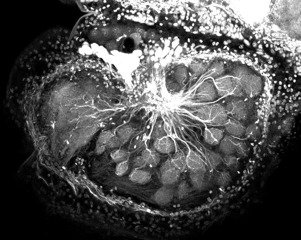
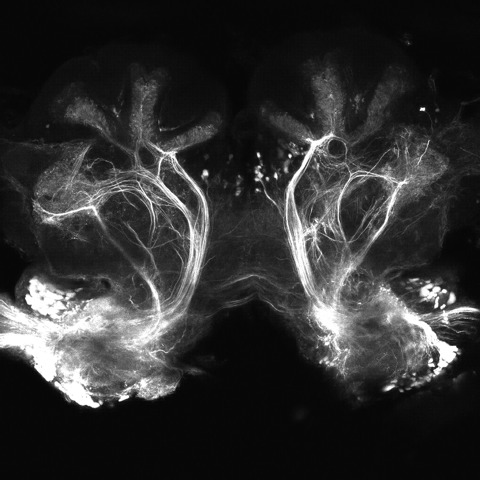
Two capillaries loaded with a chrystal of dextran-conjugated Alexa Fluor 568 were injected in both cockroach’s antennal lobes (AL). During the following 24
h, the injected dye was uptaken by AL output neurons, the projection neurons, and diffused along their axons up to their pre-synaptic terminals. Thus, while the overexposed areas identify the site of injection, it is possible to observe the projection neuron tracts leaving the AL to innervate the mushroom body and the lateral protocerebrum. The z-stack was acquired with a LSM 5 PASCAL laser scanning confocal microscope (Carl Zeiss) at a resolution of 1.16 μm/pixel (x, y) and 2 μm z-interval using a 10× dry objective (Zeiss Plan-Apochromat 10×/ 0.45). Image orientation in body axis system is bottom/ventral to top/dorsal; z-stack is from first/rostral to last/caudal.
paoli_movie1 paoli_movie1
(format.ogg)

play
download

1.5 MB

paoli_movie1
(format.avi)

play
download

17 MB

Movie 2. Z-stack of antennal lobe uniglomerular projection neurons labelling .
Antennal lobe (AL) uniglomerular projection neurons were selectively labelled by alocalized injection of dextran-conjugated Alexa Fluor 488 performed medio ventrally to the ventral mushroom body calix. During the following 24 hours of incubation, the injected dye entered the uniglomerular projection neurons (which innervate the mushroom body via the medial antennal-lobe tracts) and retro-diffused from the axons to their dendritic compartment in the AL glomeruli. Optical sections were acquired with a LSM 510 laser scanning confocal microscope (Carl Zeiss) at a resolution of 0.63 μm/pixel (x, y) and 3 μ m z-intervals using a 10× water-immersion objective (Zeiss C-Apochromat 10×/0.45w). Image orientation in body axis system is bottom/ventral to top/dorsal; z-stack is from first/rostral to last/caudal.
paoli_movie2 paoli_movie2
(format.ogg)


play
download

1.5 MB

paoli_movie2
(format.avi)

play
download

19.5 MB

Movie 3.  Antennal lobe spontaneous and stimulus-evoked activity.
Antennal lobe (AL) projection neurons were selectively labelled
for calcium imaging analysis by injecting a chrystal of dextran-conjugated Fura-2 medio ventrally to the ventral mushroom body calix. The following day, the AL was exposed to allow optical access, and projection neurons’ activity could be observed. This video shows 20 seconds of antennal lobe calcium imaging recorded before, during and after olfactory stimulation with a 100-ms pulse of 1-octanol diluted 1:100 in mineral oil (green rectangle indicate stimulus presentation). While intense activity can be observed upon stimulation, ongoing activity of weaker intensity can be observed also before and after odorant presentation. Odorant stimulation was delivered at t = 4 s and 14 s for a duration of 100 ms (note that the green rectangle lasts 250 ms to facilitate visualization). Colormap of raw data presented as ΔR 340/380. No filtering or post-processing was applied to this video. Image orientation in body axis system is bottom/ventral to top/dorsal. Scale bar, 100 μm.
paoli_movie3 paoli_movie3
(format.ogg)


play
download

5.2 MB

paoli_movie3
(format.avi)


play
download

38.5  MB