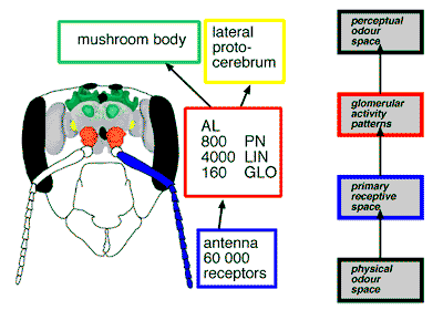
(PN: projection neurons,
LIN: local interneurons,
GLO: glomeruli)
Research group in insect olfaction
We are interested in understanding the mechanisms of the olfactory code. How do animals recognize different odors, odor mixtures and concentrations?
 |
Schematic view of the honeybee Antennal lobe. (PN: projection neurons, LIN: local interneurons, GLO: glomeruli) |
Our study is focussed on the insect antennal lobe (AL). The AL is the first central neuropil in which olfactory information is processed. It is the analogue of the olfactory bulb of vertebrates: both the AL and the bulb are structured in functional subunits, the olfactory glomeruli. These glomeruli are innervated by afferent receptor neurons, are interconnected by local, often inhibitory interneurons, and relay their processed information to higher-order brain centers. Odors are coded in across-glomeruli activity patterns which are combinatorial. That is, a glomerulus may be part of the code for quite different odors, and an odor is generally coded in the activity of a specific pattern of glomeruli, rather than a single glomerulus. Using optical imaging techniques, it is possible to simultaneously measure the activity of several glomeruli, and thus monitor their role in olfactory coding. This technique has been successfully applied to honeybees, ants, flies and moths.
We are characterizing the AL morphologically and functionally. For the honeybee the results are a morphological and a functional atlas. With the honeybee morphological atlas, each glomerulus in the AL can be identified on the basis of its shape and relative position to landmarks and other glomeruli. With the honeybee functional atlas, we document olfactory response patterns across the AL glomeruli. Likewise, we have published a morphological atlas of the glomeruli of some moth ALs, the moth morphological atlas. A morphological atlas of the fly AL (Drosophila melanogaster) has been published by other groups. We are now developing a functional atlas for the fly AL.